Proper self-examination of breasts
By way of causes of breast cancer, various environmental exposures have been investigated for associations with breast cancer.
Both active and passive exposure to tobacco smoke, charred and processed meat consumption, alcohol intake, exposure to pesticides, radiation and environmental and dietary oestrogens have all been investigated.
Of these environmental exposures, only high doses of ionising radiation to the chest area, particularly during puberty, have been unequivocally linked with an increased risk of developing breast cancer in adulthood.
Early breast cancers may be asymptomatic. Pain and discomfort are typically absent. If a lump is discovered, the following may indicate a possible presence of breast cancer:
If there is change in the size or shape of the breast.
If there are skin changes particularly dimpling of the skin over the breast lump.
If the nipple has recently inverted or has become abnormal in its shape or look.
If there is an unusual discharge, particularly blood-stained discharge from the nipple and if a lump is felt in the armpit or the tail of the breast.
For a lot of women, accurate self-examination of the breast will suffice.
Steps to aid in self-breast examination
Step 1: Begin by looking at the breasts in the mirror with the shoulders straight and arms on the hips. In the mirror, observe for unusual size, shape and colour of the breasts. Also, important is to look for unevenly shaped, distortion or swelling of either or both breasts.
Step 2: Do same as step 1, this time with the arms raised and look for the same changes.
Step 3: While at the mirror, look for any signs of fluid coming out of one or both nipples. This could be a watery, milky or yellowish fluid or in some cases frank blood from the nipples.
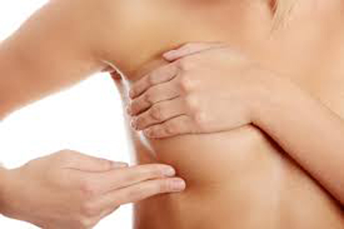
Step 4: Next, feel the breasts while lying down, using the right hand to feel the left breast and then the left hand for the right breast. Using a firm, smooth touch with the finger pads of the hand, keeping the fingers flat and together, examine the breast thoroughly. Cover the entire breast from top to bottom, side to side — from the collarbone to the top of the abdomen, and from the armpit to the cleavage. Do not forget to examine the “tail” of the breast which is the part into the armpit.
Follow a pattern to be sure that the whole breast is covered. Begin at the nipple, moving in larger and larger circles until the outer edge of the breast is reached. The fingers can also be moved up and down vertically, in rows, reminiscent of mowing a lawn.
This up-and-down approach seems to work best for most women. Be sure to feel all the tissue from the front to the back of the breasts including the tail of the breast: for the skin and tissue just beneath, use light pressure; use medium pressure for tissue in the middle of the breasts; use firm pressure for the deep tissue in the back. When the deep tissues are reached, the ribcage should be felt.
Step 5: Finally, feel the breasts while standing or sitting. Many women find that the easiest way to feel their breasts is when their skin is wet and slippery, so they like to do this step in the shower. Cover the entire breast, using the same hand movements described in step 4.
If a lump is felt, breast cancer may be present if it is hard, irregular, nodular and fixed to the skin or muscle. Any mass or suspicious findings noted on self-breast examination is an indication to get attention from a medical practitioner to confirm the findings.
Mammogram
Breast cancer is often first detected as an abnormality on a mammogram before it is felt by the patient or health care provider. Mammogram is a specialised x-ray used to examine the breast looking for early cancer. During mammogram, the breasts are compressed between two firm surfaces to spread out the breast tissues. X-rays then capture black and white images of the breast that are displayed on a computer screen and read by a specialist. Mammogram can heighten anxiety as suspicious findings may not always be cancerous.
Other imaging techniques that can be employed include ultrasound and Magnetic Resonance Imaging (MRI). Ultrasonography and MRI of the breasts are more sensitive than mammography for detecting aggressive breast cancer. Combining mammography, clinical examination and MRI is more sensitive at picking breast cancers than any other individual test or combination of tests.
Comments are closed.